

Sea Horse
A Cell Mito Stress Assay is processed at our department which contains the standard framework aimed at understanding both the physiologic and pathophysiologicfunction of mitochondria, and to predict the ability of cells to respond to stress and/or insults.
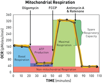
First injection:Oligomycin(ATP coupler)
Second injection:FCCP(ETC accelerator)
Third injection: Antimycin A/ Rotenone(Mito blockers
Oxigraph
Permeabilized muscle fibers can be used for subsequent analysis of oxygen consumption by an oxygraph. HRR (High Resolution Respirometry) is generally used to study:
•mitochondrial physiology and pathology
•substrate-uncoupler-inhibitor titrations, kinetics, OXPHOS flux control
•covering the entire physiological oxygen range and the extremes of hypoxia
•respiration in the closed chamber or open chamber at steady-state oxygen concentrations
Glucose substrate uptake
Glucose uptake can be analyzed by measuring the rate of uptake of radioactively labelled 2-Deoxy-D-Glucose in differentiated SAT or ASC cells. After starvation the cells will be treated with insulin (ligand binding to receptor) to trigger the translocation of glucose transporters to the cell surface. The cells subsequently are treated with a cocktail containing 2-deoxy glucose and radio-active 2 deoxy-D-(3H) –glucose. The radioactively tagged glucose will enter the cell along with the normal glucose. Measuring the labelled substrate uptake by using liquid scintillation counting provides a measure for insulin stimulated glucose uptake in the cells.
Western blotting

Proteins will be size separated via a polymerised Acrylamide/Bisacrylamide (AA/BA) gel of which the poresize is dependent on the percentage AA/BA used.The percentage of AA/BA generates the order of polymerisationof the gel. SDS is a strong anionic detergent applied to the protein sample to linearize proteins and to impart a negative charge to the linearized proteins. SDS binds proportional to most individual polypeptides, resulting in a specific charge for each polypeptide (charge per unit mass). The specifically charged polypeptides will be separated by gel migration when current is applied. By means of using a molecular weight marker, the protein size can be determined. Note: modifications of a protein, s.a. glycosylation or phosphorylation, can have a significant impact on the migration features of the protein. Due to this probably only the molecular weight can be determined.